Multiple Myeloma
& Free Light Chains
By Dr Wessel Jenner
Published September 2024
What are free light chains?
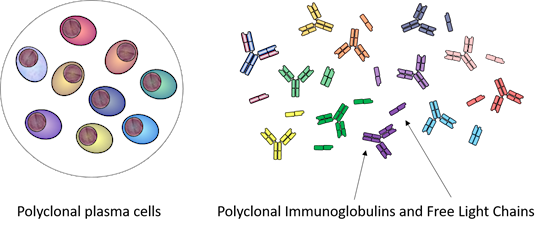
Light chains are small proteins produced in two isoforms (kappa, K; and lambda, λ) by plasma cells which are responsible for producing antibodies. Light chains are bound to heavy chains to form an antibody, also called immunoglobulin. Heavy and light chains are produced independently and assembled together within the plasma cell; however, light chains are produced in excess and secreted as free K and free λ light chains (KFLC, λFLC) in the blood (i.e., not bound to a heavy chain as it happens in an intact immunoglobulin).
When do free light chains matter?
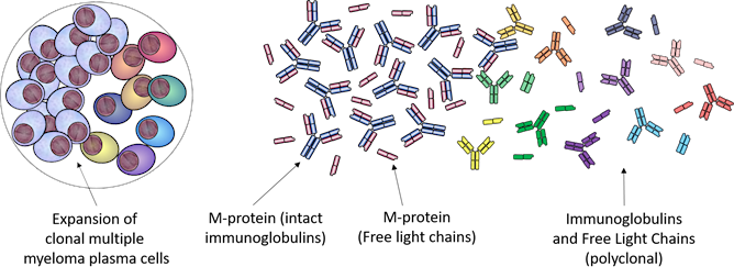
When a monoclonal gammopathy (or plasma cell dyscrasia) develops, the majority of plasma cells retain the capability of producing intact immunoglobulins and free light chains. These can be used as biomarkers in multiple myeloma. As the plasma cells grow uncontrollably from one single plasma cell clone, the blood of the patient may have increased levels of a monoclonal protein (M-protein). In the majority of myeloma patients, the M-proteins can be both intact immunoglobulins and free light chains (FLC); however, around 20% of multiple myeloma patients only secrete FLCs or secrete low levels of intact immunoglobulin.
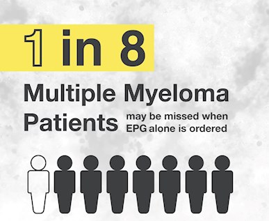
Serum protein electrophoresis (EPG) and capillary zone electrophoresis (CZE) lack the sensitivity to detect low levels of serum FLC (sFLC); in line with this, 1 in 8 patients may be missed if sFLCs are not investigated alongside EPG when ruling out multiple myeloma.1
For this reason, international2 and national3 guidelines recommend including sFLC testing when querying multiple myeloma to maximise the detection of signs indicative of myeloma. Multiple myeloma can be very difficult to diagnose due to the vague nature of its symptoms, and this is reflected in a longer time to diagnosis compared to other blood cancers.4 This is why it is pivotal that the correct tests are requested if myeloma is suspected. Through the calculation of serum KFLC / λFLC ratio, it is possible to identify plasma cell clonality and by combining EPG (and immunofixation) and sFLC analysis, >99% of myeloma patients can be correctly diagnosed.1
EPG and sFLC measurements are also used when monitoring patients with multiple myeloma and they aid clinicians in the assessment of response to therapy.5
Why is serum free light chain testing recommended over urine testing?
Free light chains are sometimes referred to as Bence-Jones Protein (BJP) when they are identified in urine by the presence of a band in urine protein electrophoresis or immunofixation. However, looking for FLCs in urine can be complicated.
Normal plasma cells produce 0.5-1 g of FLC per day; however, 10-30 g/day can be metabolised by healthy kidneys.6 This means that for FLCs to be detected in the urine, the production needs to be higher than the metabolic capacity of the kidney, or the kidney needs to be damaged. Not all myeloma patients will produce high levels of FLC or have impaired kidney function at diagnosis, and FLC might not be present in the urine, potentially leading to delays in diagnosis.
It has been demonstrated that sFLC measurement provides equivalent or better diagnostic sensitivity to urine analysis1, 8-11 in myeloma diagnosis.
If FLCs are present in the urine, they were in serum first: with just one blood sample it is possible to test both EPG and sFLC, and complying with the guideline recommendations for ruling out multiple myeloma.
If you enjoyed this article, subscribe to our electronic Pathology Focus newsletter.
References
- Katzmann JA, et al. Screening panels for detection of monoclonal gammopathies. Clin Chem 2009; 55:1517-15222.
- Rajkumar SV, et al. International Myeloma Working Group updated criteria for the diagnosis of multiple myeloma. Lancet Oncol. 2014; 15:e538-e5483.
- Quach H & Prince H. Clinical practice guideline: multiple myeloma. Australia: Myeloma Australia 2022
- Howell DA, et al. Time-to-diagnosis and symptoms of myeloma, lymphomas and leukaemias: a report from the Haematological Malignancy Research Network. BMC Hematol 2013; 13:9
- Kumar S, et al. International Myeloma Working Group consensus criteria for response and minimal residual disease assessment in multiple myeloma. Lancet Oncol 2016; 17:e328-46
- Dispenzieri A, et al. International Myeloma Working Group guidelines for serum-free light chain analysis in multiple myeloma and related disorders. Leukemia 2009; 23:215-224
- Robson EJD, et al. Utility of serum free light chain analysis when screening for lymphoproliferative disorders. Lab Med 2009; 40:325-329
- Hill PG, et al. Serum free light chains: an alternative to the urine Bence Jones proteins screening test for monoclonal gammopathies. Clin Chem 2006; 52:1743-1748
- Holding S, et al. Use of serum free light chain analysis and urine protein electrophoresis for detection of monoclonal gammopathies. Clin Chem Lab Med 2011; 49:83-88
- McTaggart MP & Kearney EM. Evidence-based use of serum protein electrophoresis in laboratory medicine. Clin Chem Lab Med 2013; 51:e113-e115
- Baulch DA, et al. Optimising the Laboratory Detection of Plasma Cell Proliferative Disorders. Presented at IBMS 2015 2015
About the Author

Dr Wessel Jenner
BSc MBChB FRCPA- Chemical Pathology
- Vitamin D Testing
- Diabetes Management
- Thyroid Function
- Hormone Testing
Dr Jenner began training in Chemical Pathology in 2001 and obtained Fellowship from the Colleges of Medicine of South Africa in 2004, as well as a Master’s degree in Chemical Pathology from the University of Pretoria in 2005. He has worked as a senior registrar in Clinical Biochemistry at the Royal Infirmary of Edinburgh, as a consultant clinical biochemist at the NHS Borders Hospital (Scotland), and as a consultant chemical pathologist in private practice in South Africa. In 2012, Dr Jenner relocated to Australia and worked as a senior registrar at the Royal Brisbane and Women’s Hospital. He obtained his Fellowship from the Royal College of Pathologists of Australasia in 2013 and joined Australian Clinical Labs in early 2014.
Related Pages
Signs & Symptoms of Multiple Myeloma
This article outlines the key blood tests for early detection of multiple myeloma to enable timely diagnosis and prevent serious complications.

QuantiFERON-TB Gold
Associate Professor Owen Harris explains the use of QuantiFERON-TB Gold assay for diagnosing latent tuberculosis and interpreting results.